|
Protocol
for RNA Quantitation Using RiboGreen™
1.
Introduction
The
Turner BioSystems TD-700 Laboratory Fluorometer in combination
with Molecular Probes’ RiboGreen™ RNA quantitation
reagent provides a method for ultrasensitive quantitation
of RNA in solution. Detecting and quantitating small
amounts of RNA is extremely important for a wide variety
of molecular biology procedures. These include measuring
yields of in vitro transcribed RNA and measuring
RNA concentrations before performing Northern blot analysis,
S1 nuclease assays, RNase protection assays, cDNA library
preparation, reverse transcription PCR and differential
display PCR.
The
most commonly used technique for measuring nucleic acid
concentration is the determination of absorbance at
260 nm (A260). The major disadvantages of the A260 method
are the large relative contribution of proteins and
free nucleotides to the signal, the inability to distinguish
between DNA and RNA, the interference caused by contaminants
commonly found in nucleic acid preparations, and the
relative insensitivity of the assay (an A260 of 0.1
corresponds to a 4 µg/mL RNA solution). The use of fluorescent
nucleic acid stains alleviates many of these problems.
The
RiboGreen RNA quantitation assay implemented with the
TD-700 Fluorometer can detect as little as 1 ng/mL RNA,
(Figure 1) exceeding the sensitivity of ethidium bromide–based
fluorometric assays(1) by 200-fold and A260 measurements
by 1000-fold. The linear quantitation range extends
over three orders of magnitude in RNA concentration,
using two dye concentrations. Using one concentration
of RiboGreen reagent and the recommended assay protocol,
researchers can quantitate 20 ng/mL to 1 µg/mL RNA.
By diluting the RiboGreen reagent 10-fold further, 1
ng/mL to 50 ng/mL RNA can be quantitated. Linearity
is maintained in the presence of several compounds commonly
found to contaminate nucleic acid preparations. Although
the RiboGreen reagent also binds to DNA, pretreatment
of mixed samples with DNase can be used to generate
an RNA-selective assay.
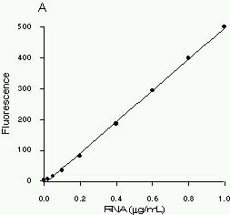 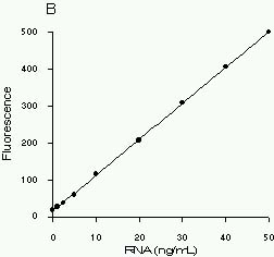
Figure 1. High-range (A) and low-range (B)
ribosomal RNA standard assays performed using RiboGreen
RNA Quantitation Reagent and the TD-700 fluorometer.
Separate instrument sensitivity calibrations were carried
out for the two assay ranges.
2.
Materials Required
- TD-700
Fluorometer with standard PMT and 10 mm x 10 mm cuvette
adaptor (P/N 7000-009).
- Blue
Mercury Vapor Lamp (10-089)
- Fluorescein
filter kit (P/N 10-086R) which includes 486 nm excitation
filter (P/N 034-0486) and 510-700nm emission filter
(P/N 10-109R-C) and two Blue Mercury Vapor lamps (P/N
10-089).
- 10
mm x 10 mm square methacrylate disposable cuvettes
(7000-959)
- RiboGreen
RNA Quantitation Kit, supplied by Molecular Probes,
Inc., Eugene, OR (catalog number R-11490). The kit
contains 1 mL of RiboGreen RNA quantitation reagent
stock solution in DMSO, 25 mL of 20X TE assay buffer
(200 mM Tris-HCl, 20 mM EDTA, pH 7.5 in DEPC (diethylpyrocarbonate)–treated
water) and 1 mL (supplied as 5 x 200 µL) of 100 µg/mL
16S and 23S ribosomal RNA standard (from E. coli),
in TE buffer. The kit contents are sufficient for
200 high-range (20 ng/mL to 1 µg/mL) RNA assays using
2.0 mL samples in 10 mm x 10 mm cuvettes. RiboGreen
RNA Quantitation Reagent (1 mL in DMSO) is also available
from Molecular Probes as a separate item (catalog
number R-11491). Handling, storage and the use of
the reagents should be performed in accordance with
the product information sheet supplied by Molecular
Probes, Inc.
- Nuclease-free
water (see Section 3.2, below)
3.
Experiment Protocol
3.1
Overview
Two different dye concentrations are required to achieve
the full linear dynamic range of the RiboGreen RNA
quantitation assay. Different working solutions of
RiboGreen reagent are prepared for the high-range
assay (20 ng/mL to 1 µg/mL RNA) and the low-range
assay (1 ng/mL to 50 ng/mL RNA), as described below
in Section 3.3.
3.2
Assay Buffer Preparation
TE assay buffer (10 mM Tris-HCl, 1 mM EDTA, pH 7.5)
is used for diluting the RiboGreen reagent and the
RNA samples. It is imperative that the TE buffer is
free of contaminating nucleases and nucleic acids.
Clean disposable gloves should be worn during handling
and preparation of all materials and solutions. All
solutions should be prepared in sterile disposable
plasticware or nuclease-free glassware, using nuclease-free
pipettes. The 20X TE buffer that is included in the
RiboGreen RNA Quantitation Kit is nuclease-free and
nucleic acid-free. This buffer is also available from
Molecular Probes, Inc. as a separate item (catalog
number T-11493). Prepare the 1X TE working solution
by diluting the concentrated buffer 20-fold with nuclease-free
water. Nuclease-free water should be prepared by treating
distilled, deionized water with 0.1% diethylpyrocarbonate
(DEPC), incubating for several hours at 37°C and autoclaving
for at least 15 minutes at 15 lbs/sq. inch to sterilize
and eliminate DEPC. Caution: DEPC is a suspected
carcinogen and should be handled with care.
Compounds containing amines, such as Tris, will react
rapidly with DEPC and should be added to DEPC treated
water only after DEPC is removed by heating. Removal
of DEPC by heating is also important to prevent carboxyethylation
of the RNA sample.(2)
3.3
Reagent Preparation
On the day of the experiment, prepare an aqueous working
solution of the RiboGreen reagent by diluting an aliquot
of the concentrated DMSO stock solution into 1X TE.
If performing the high-range assay, dilute 200-fold.
For example, to prepare enough working solution to
assay 20 samples in 2 mL volumes, add 100 µL RiboGreen
RNA quantitation reagent to 19.9 mL TE. If performing
the low-range assay, dilute 2000-fold. For example,
to prepare enough working solution to assay 20 samples
in 2 mL volumes, add 10 µL RiboGreen RNA quantitation
reagent to 20.0 mL TE. Prepare these solutions in
sterile, disposable, polypropylene plasticware rather
than glassware, as the reagent may adsorb to glass
surfaces. Protect the working solutions from light
by covering them with foil or placing them in the
dark, as the RiboGreen reagent is susceptible to photodegradation.
For best results, these solutions should be used
within a few hours of their preparation.
3.4
RNA Standard Curves
- Prepare
a 2 µg/mL solution of RNA in TE using nuclease-free
plasticware. Determine the RNA concentration on
the basis of absorbance at 260 nm (A260) in a cuvette
with a 1 cm pathlength; an A260 of 0.05 corresponds
to 2 µg/mL RNA. The 16S and 23S ribosomal RNA standard,
provided at 100 µg/mL in the RiboGreen RNA Quantitation
Kit, can simply be diluted 50-fold in TE to make
the 2 µg/mL working solution. For example, 40 µL
of the RNA standard mixed with 1.96 mL of TE will
be sufficient for the standard curve described below.
It is sometimes preferable to prepare the standard
curve with purified RNA similar to the type being
assayed. In general, equivalent amounts of single-stranded
RNA from different sources produce approximately
equal fluorescence intensity readings. The assay
remains linear in the presence of several compounds
that commonly contaminate nucleic acid preparations,
including nucleotides, salts, urea, ethanol, chloroform,
detergents, proteins and agarose. However the fluorescence
intensity may be affected (see Molecular Probes
product information sheet MP11490 for details) and
therefore the RNA solution used to prepare the standard
curve should be treated the same way as the experimental
samples and should contain similar levels of such
compounds.
- For
the high-range standard curve, dilute the 2 µg/mL
RNA solution into disposable cuvettes (or nuclease-free
plastic test tubes for transfer to quartz cuvettes)
as shown in Table 1. For the low-range standard
curve, first dilute the 2 µg/mL RNA solution 20-fold
with TE buffer to make a 100 ng/mL RNA stock solution
and use this to prepare the dilutions shown in Table
2.
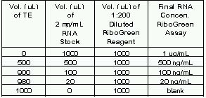
Table 1. Protocol for preparing high-range standard
curve.
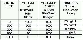
Table 2. Protocol for preparing low-range standard
curve.
- Add
1.0 mL of the appropriate aqueous working solution
of RiboGreen reagent (prepared in Section 3.3) to
each cuvette. The high-range working solution (200-fold
dilution of stock) should only be used for performing
the high-range assay and the low-range working solution
(2000-fold dilution of stock) should only be used
for performing the low-range assay. Mix well and
incubate for 2 to 5 minutes at room temperature,
protected from light.
- Set-up
the TD-700 fluorometer with a blue mercury vapor
lamp (P/N 10-089), excitation filter 034-0486, and
emission filter 10-109R-C. Calibrate the fluorometer
in simple mode according to section VII of the TD-700
manual using the sample containing the highest concentration
of RNA [Note A]. Measure the fluorescence of the
remaining samples. To equalize any photobleaching
effects, insert samples into the fluorometer for
approximately equal time periods.
- The
fluorescence reading of the reagent blank (RiboGreen
reagent + TE buffer only) may be subtracted from
that of each sample. Corrected or uncorrected data
may be used to generate a standard curve of fluorescence
versus RNA concentration.
3.5
Sample Analysis
- Dilute
each experimental RNA solution in TE to a final
volume of 1.0 mL in disposable cuvettes or test
tubes. It may be useful to prepare several dilutions
of each experimental sample. Large dilutions of
the experimental sample may serve to diminish the
interfering effect of certain contaminants. However,
extremely small sample volumes should be avoided
because they are difficult to pipet accurately.
In addition, the level of assay contaminants should
be kept as uniform as possible throughout an experiment,
to minimize sample-to-sample signal variation. For
example, if a series of RNA samples contain widely
differing salt concentrations, they cannot be compared
to a single standard curve. To avoid this problem,
simply adjust the concentration of contaminants
to be the same in all samples, if possible. See
Section 3.6 for information on eliminating DNA from
the sample.
- Add
1.0 mL of the aqueous working solution of the RiboGreen
reagent (prepared in Section 3.3) to each sample.
Incubate for 2 to 5 minutes at room temperature,
protected from light.
- Measure
the fluorescence of each sample using the same instrument
calibration conditions as used to generate the standard
curve (see 3.4.4). To equalize any photobleaching
effects, insert samples into the fluorometer for
approximately equal time periods.
- If
the standard curve has been constructed from background-subtracted
data (see 3.4.5), subtract the reagent blank fluorescence
reading from that of each of the samples.
- Determine
the RNA concentration of each sample from the standard
curve generated in Section 3.4.
3.6
Eliminating DNA from Samples
RiboGreen reagent also binds to DNA. Fluorescence
in samples that is due to RiboGreen reagent binding
to DNA can be eliminated by pre-treating the sample
with RNase-free DNase, ensuring that the entire sample
fluorescence is due to dye bound to RNA.
- Prepare
10X DNase digestion buffer: nuclease-free 200 mM
Tris-HCl, pH 7.5, containing 100 mM MgCl2 and 20
mM CaCl2.
- Add
0.11 sample volume of 10X DNase digestion buffer
to each DNA-containing sample (for example, to a
9 mL sample, add 1 mL 10X buffer).
- Add
about 5 units of RNase-free DNase I per mg of DNA
thought to be in the sample.
- Incubate
the sample at 37°C for 90 minutes.
- Dilute
the sample at least 10-fold into TE to diminish
effects of the digestion buffer salts on the RiboGreen
assay procedure.
- Perform
the RiboGreen assay as described above.
Notes
[A]. For optimal detection sensitivity, separate calibrations
should be carried out for the high range and low range
assays.
4.
References
1.
Anal Biochem 17, 100 (1966)
2. Sambrook, J., Fritsch, E.F. and Maniatis, T., Molecular
Cloning: A Laboratory Manual, Second Edition,
Cold Spring Harbor Laboratory Press (1989).
5.
Patent and Trademark Information
RiboGreen
is a trademark of Molecular Probes, Inc. RiboGreen
RNA Quantitation Reagent is covered by current or
pending U.S. and foreign patents.
|